By Dr. Rich Trenholm (Sport and Exercise Medicine)
One thing that I hear about quite from patients a bit as a Sport and Exercise Medicine physician, is that they have problem X, and they would like to have test Y to confirm what the diagnosis is.
While it is extremely important to have an accurate diagnosis for a team of rehabilitation specialists like ours at Reactivate properly target our treatment plans for our patients, not all tests are equal, and the “bigger, shinier, newer, more computerized” tests don’t necessarily mean that it is a better test.
The Best Test: The History
When we do an initial consultation with a new patient, and even at follow-ups, the first thing we do is talk. A patient will come in with a pain in the knee, for example, and then we sit down with them and talk to them for close to half an hour.
When did this issue start? Did it start suddenly? Did it creep in over time? Are some days better than others? Are mornings or evenings worse?
What activities or tasks that you do on a daily basis bother it or aggravate it? What feels good or what have you done that makes it better?
How have you changed your want of moving to compensate for the pain or issue in order to continue being active or doing your activities of daily living? What have you had to stop doing because it is too painful?
What is happening at the joints above this one that is injured? What about the one below?
And so on. What we are doing is dialing down the broad list of potential diagnoses that falls under the umbrella of “knee pain” and figuring out what two or three potential diagnoses may be. Often, but not all the time, a joint or tissue gets injured at the weakest link. So, the problem might not lie in where the injury is. Rather the problem lies somewhere else in the body that lead the injury to happen at the weakest link.
CONFIRMING WHAT WE ALREADY KNOW: THE PHYSICAL EXAM
Studies have shown that 80-90% of diagnoses can be made at this point without having to push, pull, tug, twist or touch a patients body. Then why do physicians do it? Well, it is to further narrow down the two or three potential diagnoses to one or two. Or it is to confirm what we already know from the history. In essence, we are putting our acquired information from the history through an “exam” to confirm or reject what we already know.
SO, ARE FURTHER TESTS NEEDED AT THIS POINT?
Sometimes yes, and sometimes no. If there is still a question about the diagnosis, the issue is complex, or the treatment that we think might be required (i.e. bracing, surgery, shockwave therapy, massage, physiotherapy, etc) requires an image to exactly show what is going on, then one might be ordered. However, not all injuries need an image to see what is going on.
First of all, you must remember that images like x-rays, CT scans, and MRIs are often static (not moving) and non-weightbearing pictures. Almost always, an injury or a pain results from and continues to be a problem when moving. So you need an image that is moving to see how things are moving on the inside to get an accurate picture of what is going on.
However, if the diagnosis is fairly clear after the history and physical, then an image is not required at this point. Why want weeks, and sometimes months, for an image to tell us what we already know, and delay rehabilitation efforts like chiropractic therapy, physiotherapy, and massage therapy which will be the treatment anyways. A lot of the time, images don’t change our treatment plans and exercise prescriptions for patients.
When you start rehabilitating the body, if the treatment is not going as planned or the patient is not recovering, that is additional information that makes us as rehabilitation specialists scratching our head, and maybe imaging is required at that point. So, you don’t need to order a test right off the bat in order to confirm the diagnosis before treatment starts.
WHAT YOU SEE ISN’T ALWAYS WHAT YOU HAVE
Patients are often happy when they get a test result back and there is a structural injury or abnormality, like a torn meniscus or cartilage, so they can say “Aha! That’s it! Take it out!”. Where a significant number of these findings are “incidental”, meaning that they are found but are not the cause of the pain. Sometimes they are, but not always.
Remember, the history has to withstand the examination tests that we test our theories about what the diagnosis is, and then these tests confirm or reject the shortened list of diagnoses.
Here are a couple of articles that you can read to see what I mean.
Here is an article on mensical injuries found on MRI in patients with knee pain (and another one too!)
Another article on ultrasound findings for rotator cuff tears.
If a test is required, then which one do we order and why?
MAGNETIC RESONANCE IMAGING: MRI
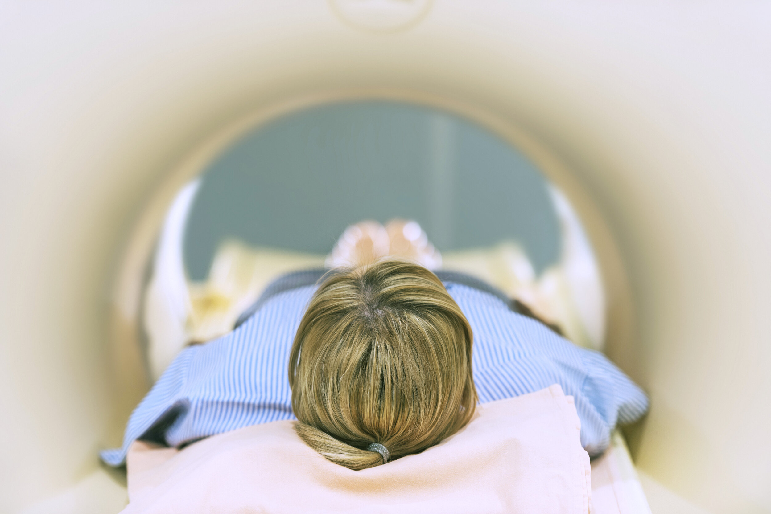
To patients, this is often thought to be the holy grail of tests. It is a big fancy machine that is loud and is run by a lot of computers. It takes weeks to months to get one of these tests, so access is not very easy. At the test, you are put onto a skinny sliding tray (so you are non-weight bearing through your joints) and you go into a tube that makes some people feel claustrophobic (they get anxious in the small space).
MRI’s are good for looking at the very small details of tissues in the body like nerves, internal ligaments, muscle fibres, and cartilage inside joints. They are also good for some subtle bone injuries, but it certainly isn’t the first thing we jump to for bone injuries.
The problem with the MRI, is that it, like other diagnostic imaging tests, is that it is non weight-bearing, and the patient isn’t moving during the test. When a patient says “Doc, it hurts when I do this…”, and then you can’t ‘do this’ during the test, then we might be able to see something. Muscles, tendons, cartilage, joints, and ligaments move and slide in your body when you move around. So you have to move sometimes to see what is going on.
CT SCAN

CT scans are more readily accessible, and often (at least here in Muskoka) we can get a CT scan within a week. The test involves another big round machine where you lie on a stretcher (non-weight bearing) and it slides into the machine that looks like the world’s biggest doughnut. It isn’t as loud as the MRI, but there is still noise. CT scans use a lot of radiation. Some more than others.
A CT of your spine is like being exposed to 6 months of background radiation that you experience on a day to day basis. A CT of a joint is much less; it is only about 3 hours of background radiation. Here is a great article on the different amounts of radiation depending on what is being scanned.
However, once again, you are non-weight bearing and not moving your body for what is often an injury or a pain that happens when you move.
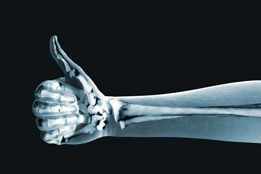
X-RAYS
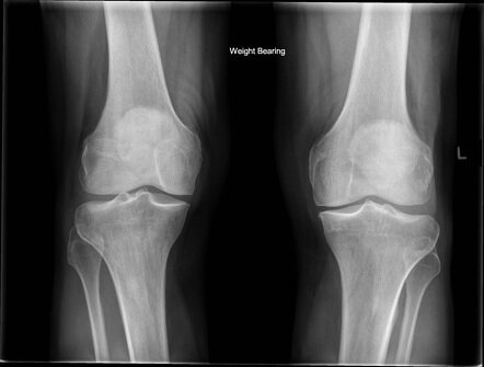
X-rays are cheap, low dose in radiation, quick, and give us a lot of information about bone and joint structure, alignment, and the nice thing is that it is weight bearing! That’s right, when you have hip, knee, ankle or foot pain that could be arthritis, we can ask for x-rays to be done while the patient is putting weight through those joints. It is amazing the difference that we see between an x-ray done on the same patient in a non-weight bearing versus weight bearing position. And it often will influence what treatments we will offer them.
Additionally, if it hurts to do something in a certain position, then we can put the patient in that position and take a picture of it.
The only downside is that x-rays are only good for bone (in the sports medicine world) injuries and joint injuries. They don’t tell us much about soft tissue injuries, but we can see certain things on x-rays that give us ideas about soft tissue injuries (like muscle, tendon, and ligaments) but we can’t see those tissues directly.
ULTRASOUNDS

Ultrasounds are undervalued and underappreciated when it comes to musculoskeletal injuries or concerns. They see soft tissues, blood flowing in the body, the surface of bones, the outer limits of joints, and the best thing is that it is a MOVING IMAGE. Yup. That’s right. Moving.
This means that when the patient says “Doc, it hurts when I dod this…”, then we can actually get them to do that when we are ultrasounding them. We can see how parts move in relation to each other. It is a functional test that gives us an incredible amount of information.
The test requires the ultrasonographer (person who does the ultrasound) to have specific and specialized training for musculoskeletal imaging, but we are very fortunate to have a few very good ultrasonographers in Muskoka. So you don’t have to drive out of town to get a test. You might need to wait a bit, but that should not stop a patient from starting their rehabilitation.
IS WAITING OK? SHOULD YOU HAVE A TEST BEFORE YOU START REHABILITATION?
Sure you can wait, but while you wait for a test, which may be weeks, muscles will atrophy (get smaller and weaker), joints will become more unstable, cartilage will further be stressed and damaged. As a comprehensive rehabilitation team, as we have at Reactivate, we work together with the best possible diagnosis at hand and treat patients while they wait for the tests.
In a future article, we will chat about the normal process of recovery and what to expect. But recovery and rehabilitation take weeks to months of consistent efforts and a progressive series of exercises to recover. So, there is no reason to wait for a test before a patient starts their rehabilitation journey.
BOTTOM LINE
Not all tests are equal, and the bigger fancier machines won’t always give your physician and rehabilitation team the information they need to get patients better. Additionally, tests aren’t always required to “see” what is going on. Most of the time we know what is going on just from talking to patients and watching them move.
So, come on into Reactivate, let us figure out what is going on with one of our sport and exercise medicine physicians (covered by OHIP with a referral from your primary care provider) and then let our team of rehabilitation specialists reactivate you so you can get back to enjoying life to its fullest.